Congenital vertical talus
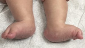
Vertical talus, also known as congenital vertical talus or CVT, is a rare deformity of the foot, typically diagnosed at birth. It presents as an extreme case of flatfoot. One or both feet can be affected.
The talus bone is the small bone in the ankle that sits between the tibia and fibula of the lower leg (shin) and the calcaneus (heel bone). The talus connects the lower leg to the foot, and it typically points towards the toes. In a foot with vertical talus, the talus points towards the ground.
With the talus positioned in the wrong direction, the bones that normally form in front of it now sit on top of the talus. The front end of the talus points down towards the sole of the foot and the bones at the end of the foot bend back upward. This results in a stiff foot with no arch that is frequently called a “rocker bottom foot”.
Vertical talus can occur by itself or may be associated with other conditions such as arthrogryposis, spina bifida, or other neurologic conditions. Because of these associations, your doctor may wish do to additional tests.
There is also a less severe form of this deformity called oblique talus. In children, with oblique talus, the talus bone is positioned in the wrong direction while weight bearing, but aligns normally when the foot is pointed down. The foot appears to be more severe than the usual flatfoot, but less severe than a foot with vertical talus.
Treatment
Vertical talus should be treated with the goal of providing a painless, stable, functional foot that fits in shoes comfortably. Initially, vertical talus is not painful. If left untreated, however, the foot will remain deformed and eventually become painful as the child begins to walk and increase activity. Calluses or skin breakdown can occur in the flattened arch area, making it difficult to wear shoes and walk.
Nonsurgical treatment
Some surgery is usually needed to correct the vertical talus deformity. Before surgery, however, your doctor may recommend a trial of stretching or casting to improve the flexibility of the foot. This may decrease the amount of surgery that is needed, or, in some cases, prevent the need for surgery all together. Casting usually requires multiple visits to your doctor since casts need to be changed almost weekly for multiple weeks. This serial casting technique allows the foot to stretch gradually without causing too much pressure in any one area at any one time.
Surgical treatment
If the foot doesn’t correct with non-surgical techniques, surgery will likely be recommended. If some of the deformity improved with casting, the surgery may be limited to behind the ankle area alone. If, however, the foot remains rigidly deformed, the surgery will be more extensive and typically done just prior to one year of age.
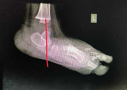
Before treatment
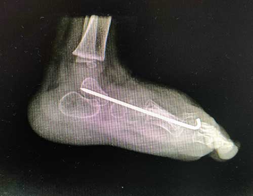
After surgery
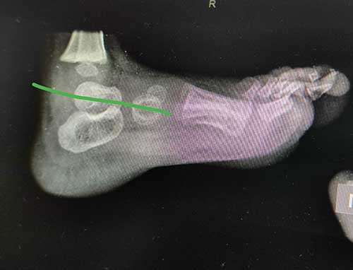
One year After surgery
The goal of surgery is to correct the position of the bones within the foot. This will likely require lengthening tendons or ligaments to allow the bones to be moved. The bones are then held in place with pins and a cast. The pins can usually be removed in the office in 4 to 6 weeks. A special shoe or brace may be recommended to try to prevent recurrence of the deformity. Your doctor will likely recommend follow up visits for a few years to see make sure the foot grows well and doesn’t need additional treatment.